


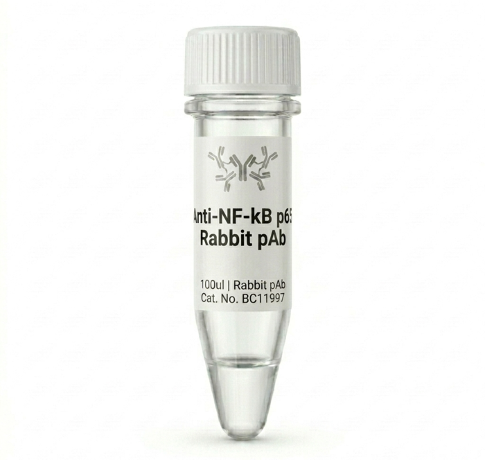
Anti-Iba1 Rabbit Polyclonal Antibody 100ul BC11105
Achieve highly specific and reliable results in your neuroscience research with our premium Anti-Iba1 Rabbit Polyclonal Antibody. Designed for superior sensitivity, this 100ul antibody is the perfect tool for identifying microglia and macrophages in fixed tissue samples. Accelerate your experimental progress with a high-affinity reagent that consistently delivers clean, reproducible staining and clear imaging.
Product successfully added to the Quote List

Description
Introduction
The Anti-Iba1 Rabbit Polyclonal Antibody is a high-performance research reagent essential for detecting Iba1, a classic marker for microglia and macrophages. Developed for accuracy and consistency, this antibody allows researchers to visualize inflammatory responses and neural processes with exceptional clarity and precision.
Key Features
– High specificity for Iba1 protein targets
– Optimized for consistent performance in standard laboratory protocols
– Provided in a convenient 100ul pack size for precise usage
– Suitable for various immunohistochemistry and immunocytochemistry applications
– Validated for reliable results in complex tissue environments
– Long shelf life when stored according to recommended conditions
Benefits
Using this Anti-Iba1 antibody streamlines your laboratory workflow by reducing the need for extensive optimization. Because it is highly specific, you will experience lower background noise and higher signal-to-noise ratios, leading to more publishable, high-quality data. It effectively solves the problem of non-specific staining, ensuring that your identification of microglial populations is accurate and statistically sound for every experiment you conduct.
Usage / How to Use
This product is intended for professional laboratory use, specifically in immunohistochemistry (IHC) or immunocytochemistry (ICC) assays. Typically, the antibody is diluted in an appropriate blocking buffer and incubated with fixed tissue sections or cell cultures to bind to the Iba1 target. After washing away unbound antibodies, a secondary antibody conjugated to a fluorophore or enzyme is used to visualize the microglia under a microscope or imaging system.
Why Choose This Product
– Trusted consistency that ensures your experiments remain reproducible across multiple trials.
– Exceptional affinity that detects even low-abundance microglial markers in difficult tissues.
– Rigorously tested for quality control to meet the high demands of modern academic and clinical research.
– Dedicated technical support and documentation to help you achieve your specific experimental goals.
Additional information
| brands | |
|---|---|
| capacity |
General Inquiries
There are no inquiries yet.

Reviews
There are no reviews yet.