
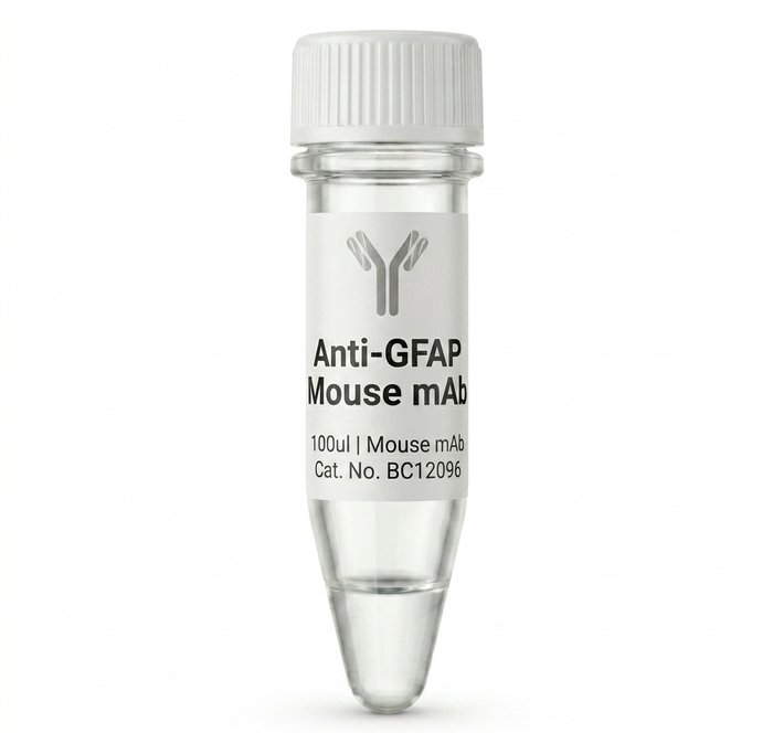
BC12096 Anti-GFAP Mouse Monoclonal Antibody 100ul
Achieve precise and reliable results in your immunohistochemistry and western blot experiments with our BC12096 Anti-GFAP Mouse Monoclonal Antibody. Designed for high specificity and sensitivity, this 100ul vial ensures consistent staining of astrocytes, making it an essential tool for neurobiology research and diagnostic applications. Trust in quality for your lab.
Product successfully added to the Quote List

Description
Introduction
The BC12096 Anti-GFAP Mouse Monoclonal Antibody is a high-performance reagent engineered for researchers focused on neurobiology and glial cell analysis. This primary antibody provides exceptional binding affinity for Glial Fibrillary Acidic Protein, allowing for clear visualization and quantification of astrocytic cells in complex biological samples.
Key Features
– High specificity for Glial Fibrillary Acidic Protein target antigens
– Optimized for immunohistochemistry and western blot protocols
– Consistent performance across multiple experimental runs
– Standardized 100ul pack size for laboratory convenience
– Validated for reliable detection in various tissue preparations
– Stable storage profile under recommended laboratory conditions
Benefits
Using the BC12096 Anti-GFAP Mouse Monoclonal Antibody significantly reduces experimental variability, saving you time and valuable reagents. By providing a clean signal with minimal background interference, this antibody allows for more accurate identification of astrocytes, which is critical for studying neuroinflammation, gliosis, and brain development. Its robust nature ensures that your data remains reproducible, strengthening the validity of your scientific findings and publications.
Usage / How to Use
This antibody is primarily used in laboratory settings for the detection of GFAP in fixed tissue sections or protein extracts. Begin by diluting the antibody according to your established protocol for immunohistochemistry or western blotting. Apply the diluted primary antibody to your membrane or tissue slide after the appropriate blocking step. Incubate as directed, then proceed with your secondary antibody detection system to visualize the GFAP-positive structures under a microscope or imaging system.
Why Choose This Product
– Unmatched sensitivity ensuring detection of even low-abundance markers
– Rigorous quality control standards for consistent batch-to-batch performance
– Backed by technical specifications that meet demanding research requirements
– Perfectly sized 100ul volume minimizes waste for routine lab tasks
– Ideal for both exploratory research and validated diagnostic workflows
Additional information
| brands | |
|---|---|
| capacity |
General Inquiries
There are no inquiries yet.






Reviews
There are no reviews yet.